


-
Recent Posts
Archives
- October 2022
- August 2022
- May 2022
- March 2022
- January 2022
- December 2021
- September 2021
- July 2021
- June 2021
- May 2021
- April 2021
- February 2021
- January 2021
- November 2020
- October 2020
- September 2020
- June 2020
- April 2020
- March 2020
- February 2020
- January 2020
- December 2019
- November 2019
- October 2019
- September 2019
- June 2019
- March 2019
- February 2019
- January 2019
- December 2018
- November 2018
- October 2018
- August 2018
- July 2018
- June 2018
- May 2018
- April 2018
- March 2018
- February 2018
- January 2018
- December 2017
- November 2017
- October 2017
- September 2017
- August 2017
- July 2017
- June 2017
- May 2017
- April 2017
- March 2017
- February 2017
- January 2017
- December 2016
- November 2016
- August 2016
- July 2016
- June 2016
- May 2016
- April 2016
- March 2016
- February 2016
- January 2016
- December 2015
- November 2015
- October 2015
- September 2015
- August 2015
- July 2015
- June 2015
- May 2015
- April 2015
- March 2015
- December 2014
- June 2014
- May 2014
- April 2014
- November 2013
- September 2013
- August 2013
- July 2013
- June 2013
- May 2013
- March 2013
- January 2013
- November 2012
- October 2012
- July 2012
- December 2011
- November 2011
- October 2011
- September 2011
- August 2011
- July 2011
- June 2011
- May 2011
- April 2011
- March 2011
- February 2011
- January 2011
- December 2010
- November 2010
Categories
Pulmonic Stenosis and Balloon Valvuloplasty in Dogs

Daisy resting comfortably at home.
Pulmonic stenosis, known as PS, is one of the most common congenital heart diseases in dogs. It can be accompanied by additional heart defects and develop into a life-threatening condition or it can be mild enough to be no more than a surprising incidental finding. Pulmonic stenosis refers to a stenosis or constriction of the right ventricular outflow tract.
Pulmonic stenosis is most commonly seen in English bulldogs like Daisy, as well as Terriers, Miniature Schnauzers, American Cocker Spaniels, Samoyeds, Keeshounds, Mastiffs and Beagles. A hereditary bases for PS has been proven in Beagles and is suspected in other breeds.
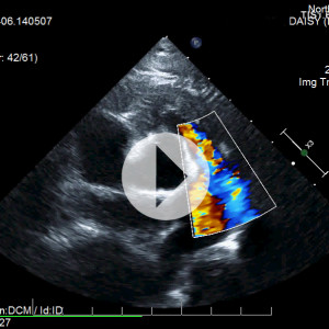 In order to understand what pulmonic stenosis is, it is necessary to understand some normal heart anatomy. The heart sits more or less centrally in the chest and is divided into a left side, which receives oxygen-rich blood from the lungs and pumps it to the rest of the body; and a right side, which receives “used” blood from the body and pumps it to the lungs to pick up fresh oxygen. Because the left side of the heart must supply blood to the whole body, its muscle is especially thick and strong while the right side, which only pumps to one nearby area, tends to be thinner. When the ventricles pump, the blood from the left shoots through the aortic valve and the blood from the right side shoots through the pulmonic valve. These valves snap sharply closed after the pumping is done. The area where the blood exits the right ventricle is called the right ventricular outflow tract and it consists of the exit area of the ventricle, the pulmonic valve, and the main pulmonary artery.
In pulmonic stenosis, the right ventricular outflow tract is narrowed either at the actual valve, just before it, or just after it. The most common form of pulmonic stenosis involves a deformed pulmonary valve such that the valve leaflets are too thick, the valve cusps are fused, or the opening is too narrow such as in the formation of an aberrant coronary artery. The heart must pump extra hard to get the blood through this unusually narrow, stiff valve. The right side of the heart becomes thick from all this extra work which may result in abnormal electrical conduction of the heart, leading to arrhythmias. In some cases these arrhythmias can be life-threatening.
A mild case is of little concern and usually does not affect life expectancy. Luckily, most cases are mild and do not require treatment; fairly severe disease is needed for clinical signs to appear.
Approximately 35-percent of dogs with severe pulmonic stenosis will show some or all of the following signs:
- Tiring easily
- Fainting spells (from the abnormal electrical heart rhythm)
- Fluid accumulation in the belly
- Blue-tinge to the gums, especially with exertion
- Difficulty breathing
- Sudden death
In some cases, medications called beta blockers can be used in an attempt to relax the muscles of the heart and open the stenosis. This will not relieve the constriction but could relieve it. In mild forms of the disease, medical management may offer the pet a good quality of life.
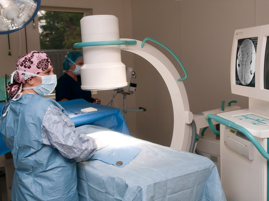
Dr. Schneiderman performing heart surgery using the C-arm at NorthStar VETS
Performing this procedure reduces the risk of sudden death by 53-percent and improves quality of life as well. However, certain types of valve deformity are not amenable to this treatment.
If your family veterinarian believes your pet needs to see a specialist, contact NorthStar VETS at 609.259.8300 to schedule an appointment with one of our veterinary specialists.
 Jennifer Schneiderman, DVM, DACVIM (Cardiology)
Dr. Schneiderman received her Doctorate of Veterinary Medicine degree at Ross University in 2009 before moving back home to Long Island, New York where she completed a one-year rotating internship in small animal medicine and surgery at Atlantic Coast Veterinary Specialists in March 2010. After that, she completed a three-year residency in Cardiology at Atlantic Coast Veterinary Specialists in July 2013 and then stayed on there as a Staff Cardiologist until June 2014. Now a board-certified Veterinary Cardiologist, Dr. Schneiderman joined the NorthStar VETS team in August 2014. Her clinical interests include treatment of congestive heart failure and complex arrhythmias along with an interest in interventional procedures such as pacemaker implantation, balloon valvuloplasty and patent ductus arteriosus occlusion. Outside of work, Dr. Schneiderman enjoys traveling, scuba diving, going to the beach and spending time with her two tuxedo cats.
The information presented on this web site is not intended to take the place of your family veterinarian’s advice and is not intended to diagnose, treat, cure or prevent any disease. Discuss this information with your own veterinarian to determine what is right for your pet. All information is intended for your general knowledge only and is not a substitute for medical advice or treatment for specific medical conditions. We can not and do not give you medical advice via this blog. The information contained in this online site and emails is presented in summary form only and intended to provide broad understanding and knowledge. The information should not be considered complete and should not be used in place of a visit, call, consultation or advice of your veterinarian. We do not recommend the self-management of your pet’s health problems.
This entry was posted in Pets, Veterinary Medicine and tagged balloon valvuloplasty in dogs, bulldogs, cardiology, congenital heart disease, jennifer schneiderman, NorthStar VETS, pulmonic stenosis. Bookmark the permalink.
Leave a Reply