


-
Recent Posts
Archives
- October 2022
- August 2022
- May 2022
- March 2022
- January 2022
- December 2021
- September 2021
- July 2021
- June 2021
- May 2021
- April 2021
- February 2021
- January 2021
- November 2020
- October 2020
- September 2020
- June 2020
- April 2020
- March 2020
- February 2020
- January 2020
- December 2019
- November 2019
- October 2019
- September 2019
- June 2019
- March 2019
- February 2019
- January 2019
- December 2018
- November 2018
- October 2018
- August 2018
- July 2018
- June 2018
- May 2018
- April 2018
- March 2018
- February 2018
- January 2018
- December 2017
- November 2017
- October 2017
- September 2017
- August 2017
- July 2017
- June 2017
- May 2017
- April 2017
- March 2017
- February 2017
- January 2017
- December 2016
- November 2016
- August 2016
- July 2016
- June 2016
- May 2016
- April 2016
- March 2016
- February 2016
- January 2016
- December 2015
- November 2015
- October 2015
- September 2015
- August 2015
- July 2015
- June 2015
- May 2015
- April 2015
- March 2015
- December 2014
- June 2014
- May 2014
- April 2014
- November 2013
- September 2013
- August 2013
- July 2013
- June 2013
- May 2013
- March 2013
- January 2013
- November 2012
- October 2012
- July 2012
- December 2011
- November 2011
- October 2011
- September 2011
- August 2011
- July 2011
- June 2011
- May 2011
- April 2011
- March 2011
- February 2011
- January 2011
- December 2010
- November 2010
Categories
NorthStar VETS Cool Case Fiona
The team at NorthStar VETS is doing innovative and amazing things every day as they work to advance the level of care available to your pet. This series of posts highlights cool cases at NorthStar VETS and the types of things done to save pets and improve their quality of life. These are cases using innovative and cutting-edge medical techniques, and/or stories of pets beating the odds. Read the story below in the doctor’s own words about the case. This is the story of Fiona, a patient of Dr. Laura Culbert of our Surgery team, and how she repaired this dog’s leg deformation.
About Fiona
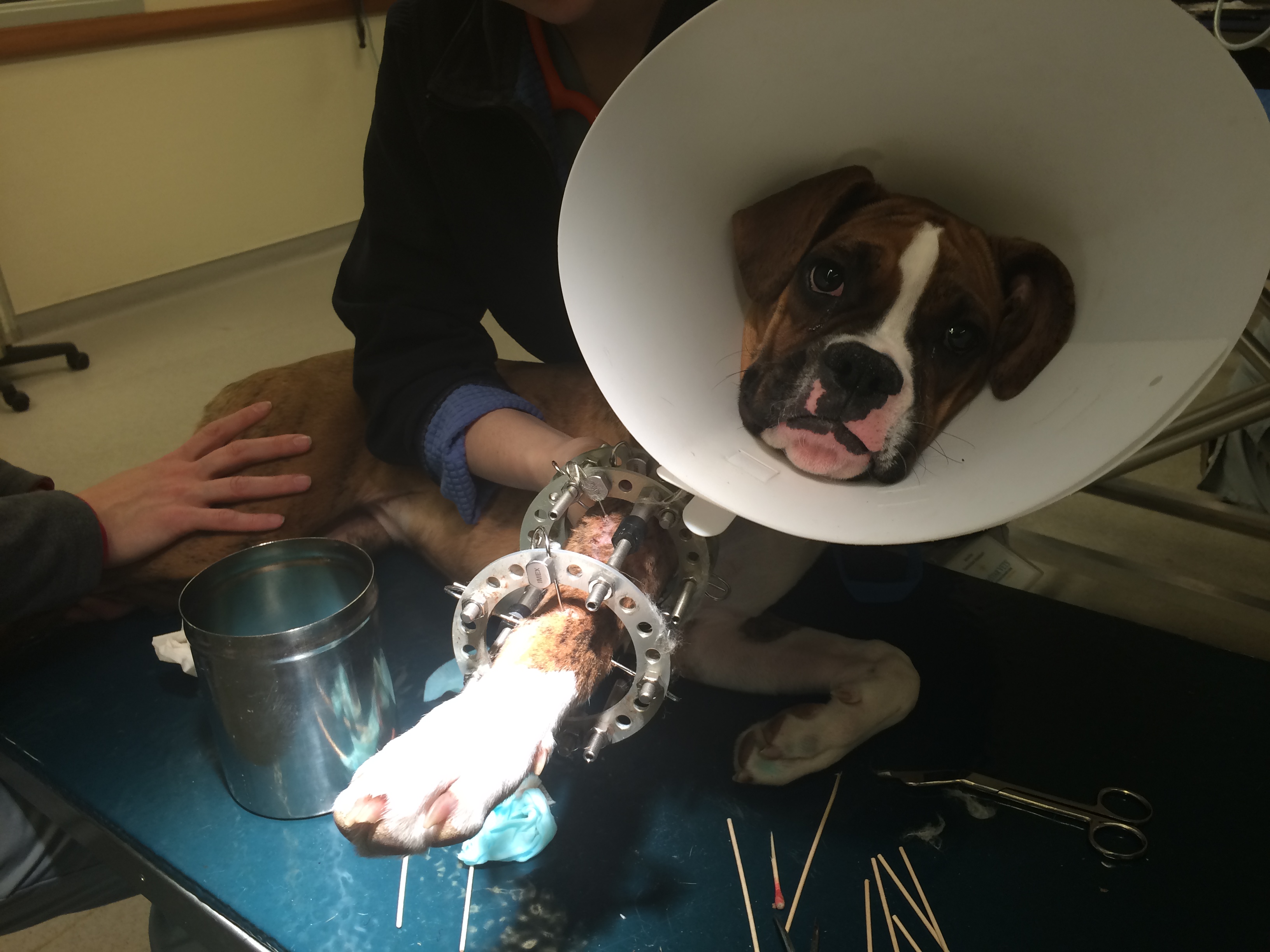 Fiona presented to the Surgery team in Robbinsville in January as a 5-month-old female intact Boxer with a right forelimb angular limb deformity. She had an abnormal curvature of the right radius and ulna which was causing her limb to curve to the inside (carpal varus). She also had a shortened right forelimb due to the premature closure of her distal medial radial growth plate.
About angular limb deformities
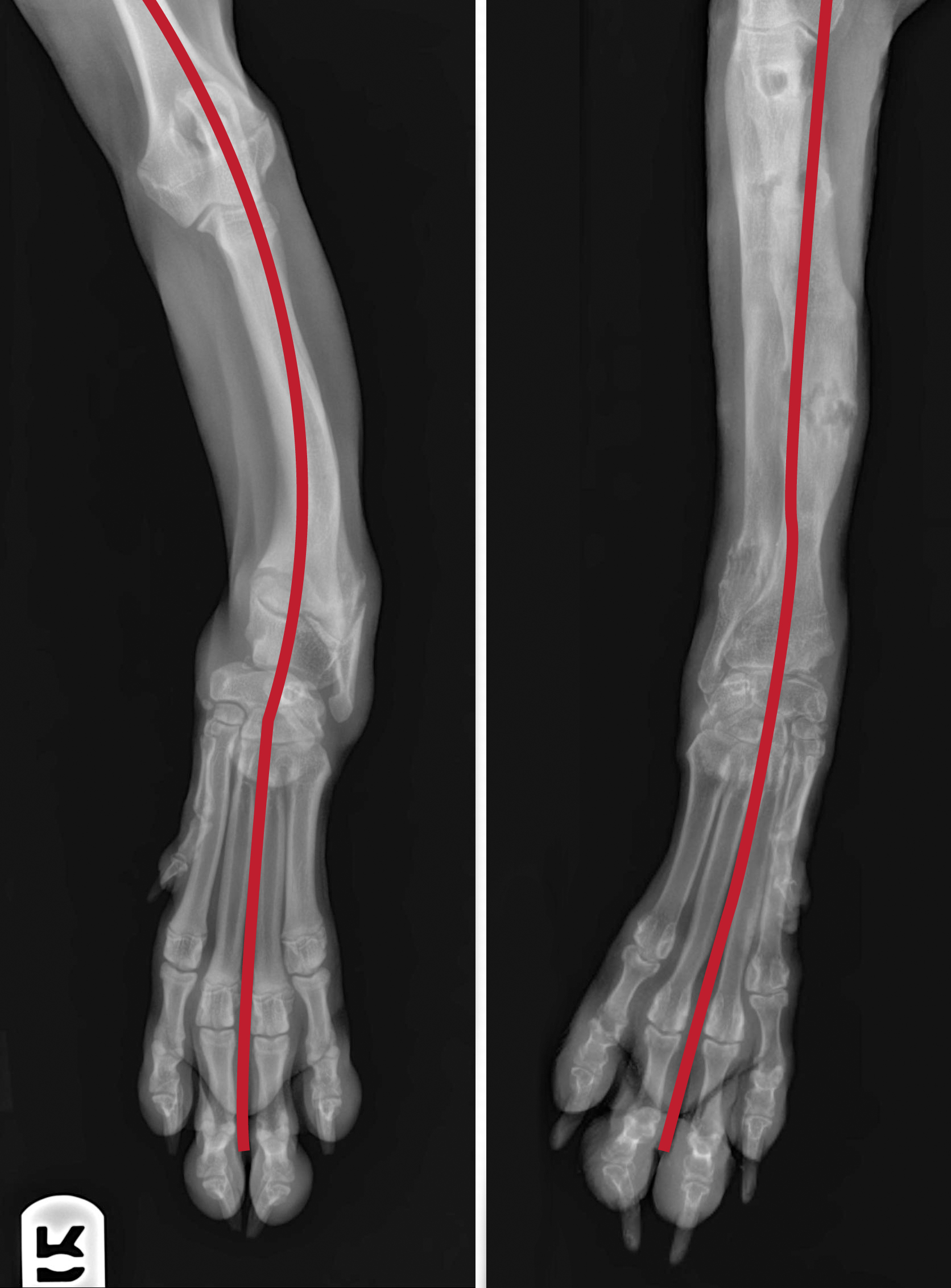
Before and after radiographs showing Fiona’s limb straightening
Angular limb deformities are challenging cases to correct because straightening the bone is of concern, but lengthening the bone is also of concern. Depending on the age of the patient and the severity of the deformity, we sometimes need to perform more than one surgery to accomplish our goals. The list of procedures to correct angular limb deformity include an ulnar osteotomy, a dome osteotomy, a closing wedge osteotomy, an opening wedge osteotomy, and distraction osteogenesis as the most common. These procedures involve cutting the bones to straighten the limb, and then procedures to lengthen the bone.
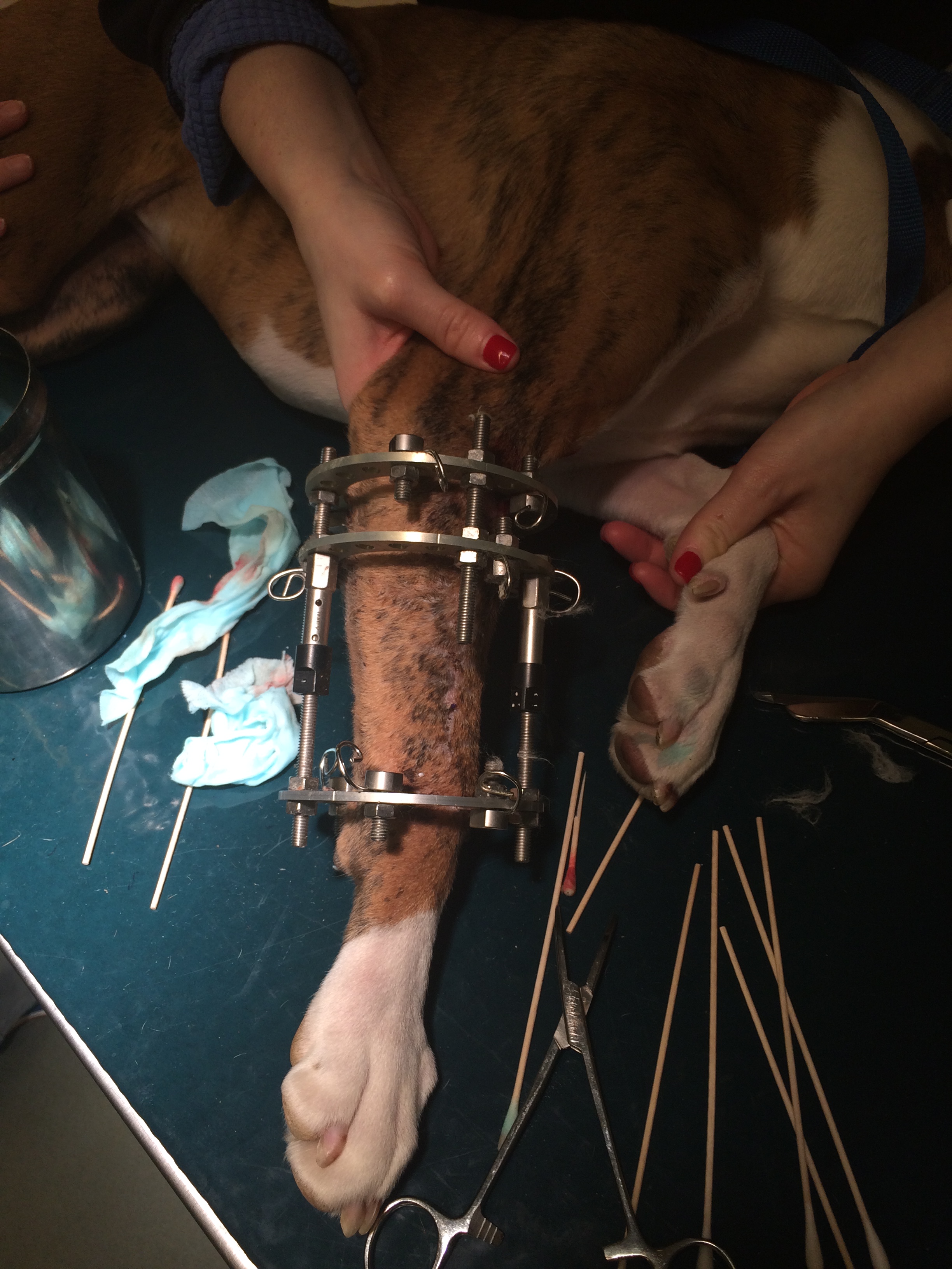
How things went for Fiona
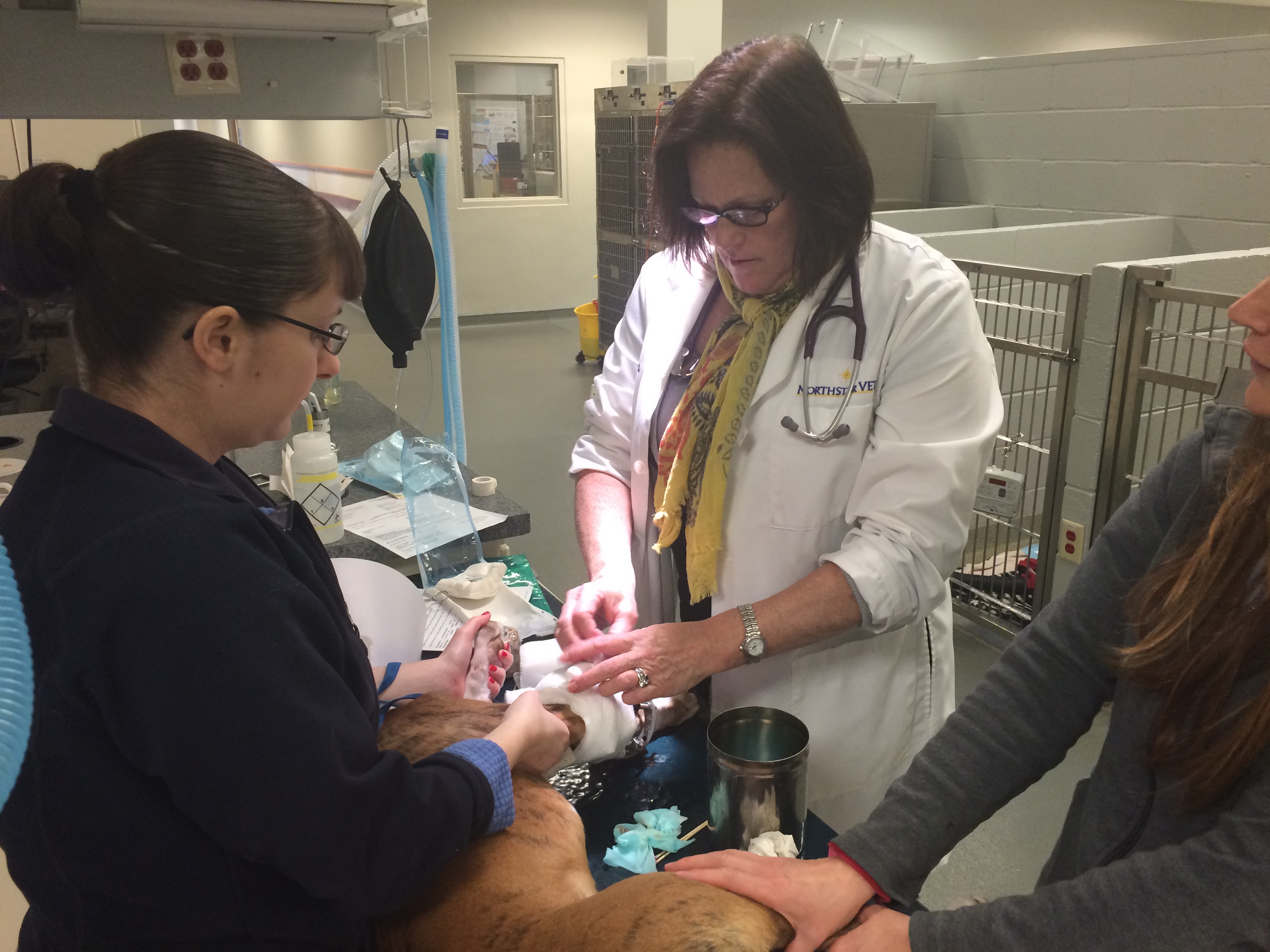
With Fiona, we performed an ulnar osteotomy, an opening wedge radial osteotomy, and distraction osteogenesis. Both radius and ulna were cut, the limb was straightened and then a circular external fixator was applied to hold the bones in place. The rings were connected with threaded rods and motors which allowed us to distract the bones across the cut each day and therefore lengthen her now straight bones.
Fiona’s parents had to follow specific instructions on how to do the distractions. What we know is that if we distract the bone at 1/4mm every 6 hours, we can achieve new bone growth that will keep up with our distractions. Therefore, a trail of bone will form in the space that we create and this bone will eventually form and harden to whatever length we need. We maintain the fixator for as long as that takes. Often the length of time in a fixator will be 8-12 weeks at minimum.
There is a tremendous amount of time put into one of these cases and a fair amount of expense. We have had a number of cases done at NorthStar VETS that have resulted in great success.
 Fiona has done very well and she now has a straight limb and a limb that is approximately the same length as her other forelimb. This allows her to be functional and pain-free. She is now 10-months old and enjoying life as an active puppy!
Learn more about the Surgery service at NorthStar VETS
 Laura Culbert, DVM, MS, DACVS
Dr. Culbert has been part of the surgical team at NorthStar VETS since 2006. She received her veterinary degree from Cornell University in 1992, and completed an internship and surgical residency at the Animal Medical Center in New York City. She has conducted research in the areas of developmental biophysiology and muscular biochemistry, and her residency project focused on neurologic diseases in dogs and complications associated with steroid therapy. Dr. Culbert’s areas of interest in veterinary surgery include cardiothoracic surgery, oncologic surgery, plastic surgery and fracture repair, and she offers the tibial tuberosity advancement (TTA) procedure for large dogs and cranial cruciate ligament repair. Dr. Culbert has worked with various rescue groups over the years including Greyhound, Australian Shepherd, Jack Russell Terrier, Golden Retriever and Boxer rescue.
The information presented on this web site is not intended to take the place of your family veterinarian’s advice and is not intended to diagnose, treat, cure or prevent any disease. Discuss this information with your own veterinarian to determine what is right for your pet. All information is intended for your general knowledge only and is not a substitute for medical advice or treatment for specific medical conditions. We can not and do not give you medical advice via this blog. The information contained in this online site and emails is presented in summary form only and intended to provide broad understanding and knowledge. The information should not be considered complete and should not be used in place of a visit, call, consultation or advice of your veterinarian. We do not recommend the self-management of your pet’s health problems.
This entry was posted in Pets, Veterinary Medicine and tagged angular limb deformity, Laura Culbert, NorthStar VETS, veterinary surgery. Bookmark the permalink.
Leave a Reply