


Specialties
Avian and ExoticsCardiology
Clinical Pathology
Dentistry and Oral Surgery
Dermatology
Emergency & Critical Care
Integrative Medicine
Internal Medicine
Medical Oncology
Neurology
Ophthalmology
Orthopedics
Radiation Oncology
Radioiodine (I-131)
Radiology
Rehabilitation & Pain Management
Surgery
Theriogenology
 Radiology
Radiology is the specialty that uses medical imaging technologies to see inside your pet's body to diagnose and sometimes treat diseases. Diagnostic imaging is an essential part of the modern veterinarian's toolkit, and at NorthStar VETS we've invested in today's most advanced imaging technology.
Radiology services are available on an outpatient basis.
Types of Diagnostic Imaging
- Radiography (X-rays)
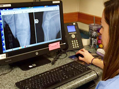
At NorthStar VETS, we use digital radiography (DR), a form of imaging in which digital sensors are used instead of traditional photographic film. A digital image-capture device records the image and makes it available as a digital file that can be viewed for interpretation, shared electronically and saved as part of your pet's medical record.
X-rays are extremely useful in detecting:
- Intestinal obstruction
- Bone fractures
- Bladder stones
- Pneumonia
- Heart disease
- Cancer
- Ultrasound
Ultrasound is another tool for evaluating your pet's internal organs. It uses sound waves that are directed into the animal and then bounce back to the ultrasound machine, forming a picture. Diagnostic ultrasound is considered the imaging modality of choice for many conditions in veterinary medicine.
While X-rays look at the outline of an organ, ultrasound tells us more about the internal appearance or architecture of the organ in a dynamic format. We often perform both radiographs and ultrasonography on a patient because the exams provide complementary information.Ultrasound is used to examine:
- Abdominal organs
- Blood vessels
- The heart
- Muscles and ligaments
- Animals in utero (in the womb)
- Ultrasound guidance can also help the radiologist perform non-invasive aspiration of internal organs and masses for more definitive diagnoses
- CT Scanning
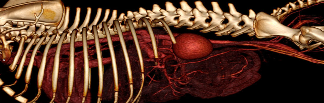
A CT (computed tomography) scan is an X-ray technique that produces images of the body's internal structures in cross-sectional "slices" rather than the two-dimensional images produced by conventional X-rays. Using a special X-ray unit that rotates around the body and a powerful computer, the CT scan produces a three-dimensional computer model of your pet's internal structures, enabling doctors to examine the body one "slice" at a time to detect disease. The contrast resolution inherent in CT far exceeds that of radiography.
Your veterinarian may recommend a CT scan to:- Diagnose muscle and bone disorders, such as bone tumors and fractures
- Pinpoint the location and margins of a tumor
- Guide procedures such as surgery, biopsy and radiation therapy
- Detect and monitor diseases such as cancer or heart disease
- Detect internal injuries and internal bleeding
- Diagnose spinal cord (herniated disc) and brain disorders
- Evaluate conditions of the nasal cavity, sinuses or inner ear
Learn how CT Angiography detected an extrahepatic portoazygous shunt in a Yorkshire Terrier.
- MR Imaging
Magnetic resonance imaging (MRI) uses a powerful magnetic field, radio frequency pulses and a computer to produce detailed pictures of organs, soft tissues, bone and virtually all other internal body structures. The images can be examined on a computer monitor, printed or copied to CD. MRI does not use ionizing radiation (X-rays).
MR imaging of the body is performed to evaluate:- Lesions of the brain and spinal cord
- Neuromuscular and musculoskeletal disorders
- Nuclear Medicine
Images using nuclear medicine technology are developed by detecting energy given off by a small amount of radioactive substance either swallowed or injected into your pet's body. As the substance travels through the body, it's absorbed by the organs and tissue and gives off what's called gamma rays. An organ that's diseased or not functioning properly emits a different energy than a healthy organ. The nuclear medicine scan "reads" this energy with a special camera and produces an image that not only shows what the organ looks like but also how it's functioning.
Nuclear medicine also offers therapeutic procedures such as Radioiodine (I-131) therapy that uses radioactive material to treat hyperthyroidism in cats.