


Specialties
Avian and ExoticsCardiology
Clinical Pathology
Dentistry and Oral Surgery
Dermatology
Emergency & Critical Care
Integrative Medicine
Internal Medicine
Medical Oncology
Neurology
Ophthalmology
Orthopedics
Radiation Oncology
Radioiodine (I-131)
Radiology
Rehabilitation & Pain Management
Surgery
Theriogenology
Ureteral Stent Case Study
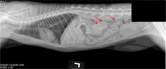
The ureter is the tube that connects each kidney to the bladder, acting as passageway for urine flow from one organ to the other. Cats and dogs can occasionally form stones or strictures that block one or both of the ureters and prevent urine from flowing. This can result in abdominal pain, and in some patients, an acute increase in their kidney values. Patients affected by complete or partial ureteral blockage may show signs of vomiting, decreased appetite, lethargy, and other signs of illness. Abdominal ultrasound and radiographs aid in the diagnosis of this condition. Below is a radiograph of a cat with stones in the ureter.
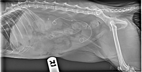
If the obstruction is not relieved, there can be a complete loss of function in that kidney. If caught early enough, a surgical procedure can be performed in which a stent is placed in the ureter. The stent spans from the kidney into the bladder, bypassing any ureteral stones present. It is hollow inside so that urine can flow both through and around the stent. Once urine flow is restored, the kidney has the potential to return to its previous level of function. Below is a radiograph of a cat after the stent procedure. You can see the white line with the two curls on each end. One curl of the stent is anchored in the kidney and the other curl is anchored in the bladder so it doesn't move.
There are risks with stent placement both short term and long-term. Those are discussed prior to the procedure. The procedure is performed by our interventional radiology team. They are always available to discuss your pet with your primary veterinarian to decide if they would be a candidate for this procedure.