

-
Recent Posts
Archives
- October 2022
- August 2022
- May 2022
- March 2022
- January 2022
- December 2021
- September 2021
- July 2021
- June 2021
- May 2021
- April 2021
- February 2021
- January 2021
- November 2020
- October 2020
- September 2020
- June 2020
- April 2020
- March 2020
- February 2020
- January 2020
- December 2019
- November 2019
- October 2019
- September 2019
- June 2019
- March 2019
- February 2019
- January 2019
- December 2018
- November 2018
- October 2018
- August 2018
- July 2018
- June 2018
- May 2018
- April 2018
- March 2018
- February 2018
- January 2018
- December 2017
- November 2017
- October 2017
- September 2017
- August 2017
- July 2017
- June 2017
- May 2017
- April 2017
- March 2017
- February 2017
- January 2017
- December 2016
- November 2016
- August 2016
- July 2016
- June 2016
- May 2016
- April 2016
- March 2016
- February 2016
- January 2016
- December 2015
- November 2015
- October 2015
- September 2015
- August 2015
- July 2015
- June 2015
- May 2015
- April 2015
- March 2015
- December 2014
- June 2014
- May 2014
- April 2014
- November 2013
- September 2013
- August 2013
- July 2013
- June 2013
- May 2013
- March 2013
- January 2013
- November 2012
- October 2012
- July 2012
- December 2011
- November 2011
- October 2011
- September 2011
- August 2011
- July 2011
- June 2011
- May 2011
- April 2011
- March 2011
- February 2011
- January 2011
- December 2010
- November 2010
Categories
NorthStar VETS Cool Case Dobby
Dobby is a Chihuahua born with deformities in his upper legs. Through advanced veterinary medicine, Dr. Daniel Stobie of the NorthStar VETS Surgical team used 3D printing as a tool in helping this dog have good legs to stand on.
Dobby had trouble walking normally as a puppy, and as he grew, the problems became more pronounced. His mom, Lori, shared her experience. “By the time he hit about a year old, we realized there was something seriously wrong with the left leg, not realizing there was anything wrong with the right leg.” Doctors soon determined just how serious Dobby’s problem was.
“His patellas were not sitting in place.” explained Dr. Daniel Stobie of the veterinary Surgery team at NorthStar VETS, “The patella is the knee cap, and normally it should sit in the middle of the femur on the leg, but it was moving off to the inside of the leg. That did not allow for him to support weight on his hind legs, and it also caused the bone in his left femur to grow crooked.”
Dr. Stobie decided to perform multiple surgeries to help fix both of Dobby’s legs. “The left leg was much worse than the right leg, the left leg was a grade 4 patellar luxation and the right leg was a grade 3 patellar luxation. We decided to repair the one that was not as badly affected first to give him a good leg to stand on after the surgery on the left side. The left leg is going to require what is called a corrective osteotomy because of the abnormal forces. What that means is that the bone grew crooked. We were able to do a CT scan of his whole pelvis and 3D print the bone. The bone has grown to where it’s sloping medially (medial varus), and is not straight because of the pressure from the patellar luxation.”
Lori commented on how things went during the surgical process. “Dobby sailed through. Everyone here is so compassionate and kind and patient with him. They always told me what to expect, told me how to handle it, and it all went as expected.”
Dr. Stobie and the team at NorthStar VETS printed a 3D model of Dobby’s bones to help plan the details of the surgery and he walked us through that process. “It allows us to look at the bone in three dimensions and measure the angles and the slope of the bone. These models are made out of a plastic that we can cut which will allow us to practice the surgery ahead of time so we can make sure we have the right angle, take the right amount of bone out, and have a special plate made that attaches to the outside of the bone to hold it in its new position once it’s repaired. You can even see from this model how far off the bone is from the straight edge of the plate.” He gestured to the 3D model held against the plate. “That degree of angulation is going to be taken out so that when we repair it, that bone will sit against the plate and be held in its new position. It’ll allow him to have a nice straight leg to walk on and should allow for us to get the knee cap back in place.”
After eight weeks of recovery after Dobby’s first successful surgery, Dobby underwent another surgery to great success. Lori gave us an update after the second surgery. “Now that he is about 90 days out after the surgery, he’s putting his leg down. Not that I doubted it would happen, but after seeing him hold it up for such a long time, it was hard to imagine how he would ever walk on four legs, but he is putting weight on it! He’s almost like a normal little dog again. In fact, he comes to work with me every day, and now instead of having to carry him everywhere we go, I have to remember to put him down on the ground and let him walk like a normal dog because he can. Everyone who sees him is actually impressed and amazed to see that he can actually walk now, because for such a long time, just to get ten feet across the room was exhausting to him. I was concerned about his back and having long-term issues with his back or other legs, but now he’s like a normal little dog. We’re really happy about it!”
Dr. Stobie and the team at NorthStar VETS will continue to utilize new technologies to better serve and treat their patients. Dr. Stobie summed up Dobby’s case. “Being able to have this new technology allows us to do new, innovative procedures. That is always exciting! Bringing the latest, greatest technology to our hospital means better patient care and better outcomes for the pets.”
Learn more about the Surgery service at NorthStar VETS along with more about the Surgery team.
Daniel Stobie, DVM, MS, DACVS – Chief of Staff
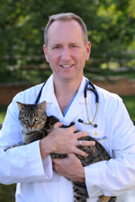 A New Jersey native, Dr. Stobie completed his undergraduate work at Cook College/Rutgers University and is a 1990 cum laude graduate of the University of Missouri-College of Veterinary Medicine. He completed an internship in small-animal medicine and surgery at the Angell Memorial Animal Hospital in Boston, then went on to complete a three-year surgical residency at the University of Minnesota and earn a Master’s Degree in Veterinary Surgery, Radiology, and Anesthesia in 1994. Dr. Stobie became a Diplomate of the American College of Veterinary Surgeons in 1995. In 2007, he completed the mini-MBA certificate program at the Rutgers School of Business. Learn more about Dr. Daniel Stobie.
This entry was posted in Pets, Veterinary Medicine and tagged 3D printing, Daniel Stobie DVM, NorthStar VETS, patellar luxation, veterinary surgery. Bookmark the permalink.
Leave a Reply